

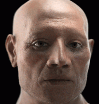
Archaeologists and forensic specialists have reconstructed the face and brain of an ancient Egyptian mummy at the Egyptian Museum in Turin. The 3,500-year-old mummy was discovered in 1904 by Italian Egyptologist Ernesto Schiaparelli, but it became really famous about two years ago when researchers identified he was the oldest case of chronic heart failure.
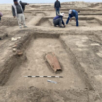
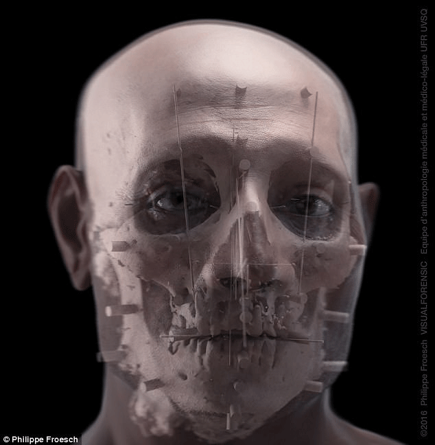
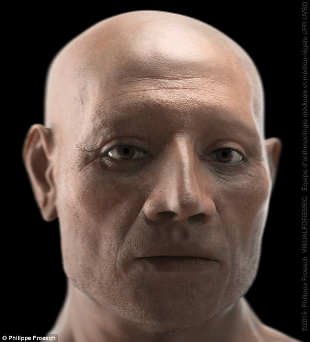
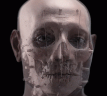
The remains belong to an Egyptian dignitary, Nebiri, who lived during the 18th Dynasty period and worked as Chief of Stables under the reign of Thutmoses III. His tomb, which was in the Valley of the Queens in Luxor, had been looted in the past and his body destroyed, but his head and canopic jars with his organs were saved. Now, modern non-invasive forensic techniques have allowed scientists to reconstruct Nebiri’s face and brain, with the use of a computed tomography and facial reconstruction techniques.
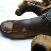
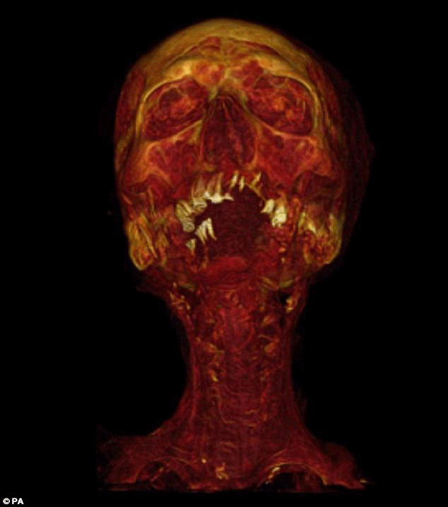
The reconstruction is based on actual anatomical features of the remains thanks to the technique used to preserve the face and skull by ancient Egyptians. The linen bandages used had been treated with a mixture of an animal fat or plant oil, a balsam or aromatic plant, a coniferous resin and heated Pistacia resin. Apart from the chemical analyses which identified the embalming materials used, CT scans showed how the bandages had been carefully inserted almost everywhere in the head. Thus, the body was protected from insect colonization allowing the facial and neck features to maintain their appearance. The scans also showed that a tiny hole had been made to the cribriform plate, a bone structure separating the nasal cavity from the brain, to insert the linen packing.
Using the data acquired from the CT scan researchers were able to produce a 3D reconstruction of the brain surface. The sophisticated method of funerary treatment helped scientists confirm the argument that Nebiri was a member of high elite.
The paper presenting the reconstruction was published in the journal Forensic Science, Medicine and Pathology.